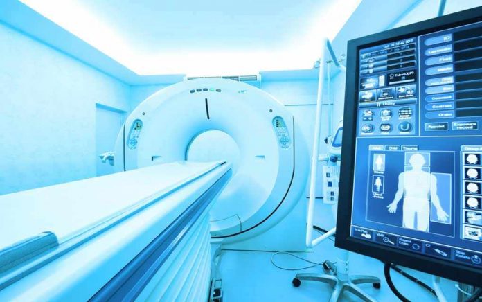
Like all medical procedures, computed tomography (CT), fluoroscopy, and nuclear medicine imaging exams present both benefits and risks. These types of imaging procedures have led to improvements in the diagnosis and treatment of numerous medical conditions. At the same time, these types of exams expose patients to ionizing radiation, which may elevate a person’s lifetime risk of developing cancer. As part of a balanced public health approach, the U.S. Food and Drug Administration (FDA) seeks to support the benefits of these medical imaging exams while minimizing the risks.
In 2010, FDA’s Center for Devices and Radiological Health (CDRH) launched an Initiative to Reduce Unnecessary Radiation Exposure from Medical Imaging and held a public meeting on Device Improvements to Reduce Unnecessary Radiation Exposure from Medical Imaging (March 30-31, 2010). These efforts were in response to increasing exposure to ionizing radiation from medical imaging highlighted in the National Council on Radiation Protection and Measurements Report No. 160External Link Disclaimer and safety concerns highlighted in FDA’s Safety Investigation of CT Brain Perfusion Scans.
Through this initiative, the FDA strives to promote patient safety through two principles of radiation protection developed by the International Commission on Radiological Protection
- Justification: The imaging procedure should be judged to do more good than harm to the individual patient. Therefore, all examinations using ionizing radiation should be performed only when necessary to answer a medical question, help treat a disease, or guide a procedure. The clinical indication and patient medical history should be carefully considered before referring a patient for any imaging examination.
- Dose Optimization: Medical imaging examinations should use techniques that are adjusted to administer the lowest radiation dose that yields an image quality adequate for diagnosis or intervention (i.e., radiation doses should be “As Low as Reasonably Achievable”). The technique factors used should be chosen based on the clinical indication, patient size, and anatomical area scanned, and the equipment should be properly maintained and tested.
FDA is pursuing efforts using its regulatory authority as it applies to imaging equipment and manufacturers. Equally important gains will be made through key partnerships with professional organizations, industry and other governmental agencies to incorporate radiation protection principles into facility quality assurance and personnel credentialing and training requirements.
Many efforts have been undertaken by a large number of groups to help ensure that each patient is getting the right imaging exam, at the right time, with the right radiation dose. FDA hopes to provide a comprehensive approach for this effort with collaborative activities in the following areas:
- Facility guidelines and personnel qualifications
- Education and communication
- Appropriate use
- Equipment safety features
- Tracking radiation safety metrics
- Research and development
Each of these areas requires coordinated efforts by regulatory, professional and industry partners to achieve common goals as described below.
Facility guidelines and personnel qualifications: For facilities participating in the Medicare program, the Centers for Medicare & Medicaid Services (CMS) has established minimum standards for hospital radiologic services, and accreditation requirements for freestanding advanced diagnostic imaging facilities. States and/or accreditation organizations may have additional requirements that go beyond the CMS requirements. In complying with these requirements, facilities can ensure adoption of policies and procedures that govern safe administration of radiation for imaging purposes. Facility accreditation can also have a direct impact on how equipment operators establish and maintain qualifications necessary to understand equipment functionality and operate equipment safely.
Education and communication: Qualified medical professionals should have ready access to up-to-date radiation safety training material, in particular for the equipment models in use at the facility. In addition, patients should have access to information that permits discussion with their medical professional on why they need a particular exam, what are the risks, and whether an alternative exam is possible. Medical professionals should be prepared to address possible patient concerns.
Appropriate use: Providers should receive training on the principle of justification and the availability of medical specialty guidelines to help assess the need for a particular exam and promote ordering of only those exams that are appropriate for the patient’s condition. In addition, automated decision support systems should be considered for implementation if data from test programs, such as the ongoing CMS Medicare Imaging Demonstration, support their use. Electronic health records should include complete information on the patient’s imaging history to aid the physician in choosing an appropriate exam.
Equipment safety features: Once an appropriate exam is ordered, the imaging equipment used should be capable of providing information and tools to operators that promote optimized delivery of radiation. Equipment features should address capture of patient information and dose, transmission of that information to data systems, controlling user access to equipment settings and features, and alerting the operator when patient safety is at risk.
Tracking radiation safety metrics: Development of information systems and analysis tools to track radiation safety metrics will play an important role in promoting radiation protection and patient safety. Collection of equipment parameters and dose for imaging exams in dose registries can be used to benchmark imaging practice through establishing diagnostic reference levels, thus improving the practice of radiology through quality assurance. A long-term goal is automated real-time updating of dose registries to facilitate comparison of exam parameters and dose indices with established reference levels, enabling immediate notification and mitigation of patient safety hazards. Tracking adverse events can establish trends and allow prospective correction of possible radiation safety problems related to equipment or operator training. Automated tracking of radiation safety metrics (e.g., through participation in a dose registry) will help fulfill quality assurance and quality improvement requirements for facility accreditation and personnel continuing education, while ensuring that operators use equipment optimally to promote patient safety.
Research and development: Continued research in medical imaging, with a focus on radiation safety and dose optimization, is crucial for all of the above areas. Scientific research facilitates better evaluation of the risks and benefits of imaging exams, and forms the foundation for the development of national and international standards for dose and image quality assessment for medical imaging devices.


